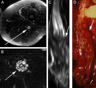
Dr. Aaron Filler's pulse-sequence innovation, which created modern MR neurography, had its first major test in November 1992, using a Signa general clinical magnetic resonance scanner. The scan below highlights the sciatic nerve structure while all other surrounding tissues stay dark. This was the first image that demonstrated the ability to capture the fascile pattern within the nerve; It was also the first image to prove that the highlighted structure was indeed a nerve.
Caption
A - Thigh cross section. The arrow indicates the sciatic nerve. This patient had suffered a stab wound to the thigh that had severed his sciatic nerve.
B - Expanded view of the sciatic nerve revealing the internal structure of the fascicle.
C - Three-dimensional reconstruction of the nerve based on the "maximum intensity projection" of the nerve. This image closely matches the shape of the sciatic nerve seen in the resonance image.
D - Intraoperative photograph taken during surgery. The photo shows the severed sciatic nerve. The arrow indicates the suture line where the grafts of
nerve were seeded in place.